Why Dental X-Rays Matter During Your First Visit

July 7, 2025
Dr. Harry Gulati, Throughout his career, Dr. G has been recognized for his exceptional work. He received the 40 under 40 award from Incisal Edge magazine and the Doctor’s Choice Award. Dr. G and White Mountain Dental have also been proud recipients of the Best of Mt. Washington Reader’s Choice Award. As a fellow of the International College of Dentists and the International Academy of Dento-Facial Esthetics (IDFE), Dr. G demonstrates his unwavering commitment to excellence in dentistry. With over a decade of experience and extensive continuing education, Dr. Gulati possesses comprehensive expertise in restorative dentistry, including crowns, bridges, implants, and oral surgery.
Introduction to Dental X-Rays
Your very first visit to Androscoggin Valley Dental sets the tone for your long-term oral health journey. One of the most critical steps during this visit is taking dental X-rays. These images serve as a diagnostic roadmap, revealing areas hidden from the naked eye, like the foundation beneath your smile. Understanding why dental X-rays matter empowers you to make informed decisions and ensures your treatment plan is both safe and effective.
What Are Dental X-Rays?
Dental X-rays are low-dose radiographic images that capture the structure of your teeth, bones, and surrounding tissues. There are several types:
- Bitewing X-Rays: Show upper and lower teeth in a single view, ideal for detecting interproximal cavities.
- Periapical X-Rays: Focus on one or two teeth from crown to root tip, helping identify root infections or bone loss.
- Panoramic X-Rays: Provide a full-mouth image in one shot, useful for evaluating jaw joints, wisdom teeth, and overall bone structure.
By integrating these modalities, your dentist gains a complete portrait of your oral health.
Why Dental X-Rays Matter on Your First Visit
1. Early Detection of Hidden Issues
Standard visual exams and probes can miss problems lurking beneath the surface. X-rays reveal:
- Interproximal Cavities: Early decay between teeth that won’t show up until it’s much larger.
- Impacted or Malpositioned Teeth: Wisdom or other teeth that haven’t erupted properly, which can crowd or damage adjacent teeth.
- Pathologic Lesions: Cysts, tumors, or other unusual growths in bone that could progress if left unchecked.
- Bone Loss & Periodontal Pockets: The first signs of gum disease, often painless until advanced.
Catching these issues before you feel pain or see symptoms means treatment can be conservative, comfortable, and cost-effective.
2. Customized Treatment Planning
Every mouth is unique, your bone density, root anatomy, and tooth position create a blueprint for care. X-ray insights allow us to:
- Map Restorations Precisely: Determine ideal crown lengths and shapes, ensuring a perfect fit and long-term seal.
- Plan Surgical Procedures Safely: From extractions to implant placements, we know exactly where nerves and sinuses lie.
- Sequence Complex Care: For patients needing multiple procedures, X-rays guide the order and timing, minimizing discomfort and overall visits.
This level of detail transforms a “one-size-fits-all” approach into a truly personalized plan designed just for you.
3. Baseline Records for Long-Term Monitoring
Your first set of X-rays becomes a trusted benchmark. As you return over the years, these images help us to:
- Track Progressive Changes: Subtle shifts in bone levels, enamel wear, or recurring decay become immediately apparent.
- Optimize Preventive Care: If we spot early thinning of enamel or widening of periodontal gaps, we can intensify cleanings or recommend targeted home routines.
- Document Treatment Outcomes: After an implant, root canal, or orthodontic adjustment, follow-up X-rays confirm that healing is on track.
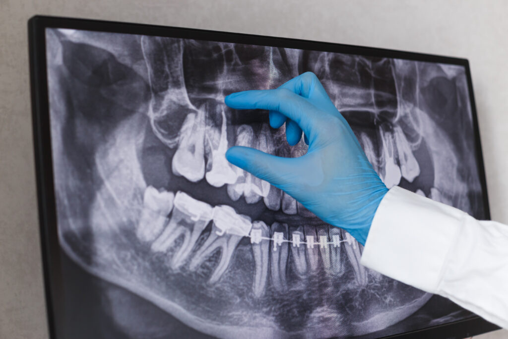
The Dental X-Ray Process at Androscoggin Valley Dental
Dental X-rays for new patients with Dr. Harry Gulati at Androscoggin Valley Dental
- Step 1: Patient Preparation
We’ll explain the process, answer any questions, and fit you with a lead apron to minimize exposure. - Step 2: Image Capture
Using state-of-the-art digital sensors, we take the necessary bitewing, periapical, and/or panoramic images in minutes, eliminating uncomfortable molds. - Step 3: Image Analysis
Within seconds, your X-rays appear on our chairside monitor. Dr. Gulati reviews them in detail, looking for areas of concern. - Step 4: Collaborative Review
We walk you through your images, highlighting findings and next steps.
This streamlined process, Dr. Harry Gulati’s dental X-ray process for new patients, ensures comfort, safety, and transparency every step of the way.
Dr. Harry Gulati’s Approach to Dental X-Rays for First Visits
At Androscoggin Valley Dental, we prioritize patient education. Dr. Harry Gulati’s approach to dental X-rays for first visits emphasizes:
- Minimal Radiation: Leveraging digital radiography, we reduce radiation exposure by up to 80% compared to film.
- Targeted Imaging: Rather than taking a full set of images on every patient, we customize the number and type of films based on your oral history and risk factors.
- Comfort-First Protocols: Rapid image capture and ergonomic positioning eliminate discomfort, even for patients with limited mouth opening.
Benefits of Dental X-Rays During the First Visit
- Comprehensive Risk Assessment: Identifies decay, infections, or abnormalities before symptoms arise.
- Enhanced Safety: Early detection of underlying issues prevents emergency procedures later.
- Informed Consent: You see exactly what we see, empowering you to participate in care decisions.
- Cost-Effectiveness: Treating small cavities or early bone loss is far less costly than addressing advanced disease.
According to a June 10, 2025 USA Today article, “It’s stone fruit season! A dietitian’s favorite ways to eat them this summer,” stone fruits like peaches, nectarines, and plums offer juicy layers beneath a firm exterior. Just as you savor the sweet interior only after peeling away the surface, dental X-rays reveal hidden layers beneath enamel, like early decay or impacted teeth, that you can’t detect by sight alone. Incorporating stone fruits into your diet boosts vitamin C and fiber, supporting gum and overall health. Similarly, routine X-rays at your first visit nurture long-term oral health by uncovering hidden concerns, ensuring your smile remains as vibrant as summer’s juiciest peach.
Considerations & Safety
- Radiation Exposure: Modern digital X-rays emit very low radiation, comparable to a few days of natural background exposure. Lead aprons and thyroid collars add an extra safety layer.
- Frequency of Imaging: We follow ALARA (“As Low As Reasonably Achievable”) principles, only recommending X-rays when clinically necessary, not by routine schedule alone.
- Child vs. Adult Protocols: Growing children may require more frequent monitoring, whereas adults with stable oral health may need fewer films.
Why Dr. Harry Gulati Uses Dental X-Rays for New Patients
By integrating radiographs into every initial exam, Dr. Harry Gulati uses dental X-rays for new patients to:
- Detecting hidden cavities before they cause pain.
- Assess bone levels for periodontal safety.
- Plan for dental or implant needs with precision.
This commitment underpins our patient-centered philosophy: prevention first, intervention when necessary.
The Future of Dental Imaging
- Artificial Intelligence (AI): Automated lesion detection may soon flag decay or abnormalities within seconds.
- 3D Printing Integration: CBCT data will allow on-site fabrication of surgical guides for implants.
- Wearable Sensors: Emerging research explores smart mouthguards that detect bruxism or acid levels in real time.
As technology advances, the clarity and utility of dental imaging will only grow, further enhancing early diagnosis and personalized care.
Key Takeaways
- Early Detection: X-rays spot decay and disease before symptoms appear.
- Customized Care: Detailed images inform precise, personalized treatment plans.
- Safety First: Modern digital radiography minimizes radiation exposure.
- Cost Savings: Treating early issues prevents complex, costly procedures later.
- Baseline Records: Initial X-rays serve as reference points for your long-term oral health.
FAQs
1. How often do I need dental X-rays after my first visit?
Frequency depends on your individual risk factors, patients with a history of cavities or gum disease may need X-rays every 6–12 months, while low-risk adults might be imaged every 24–36 months.
2. Are dental X-rays safe during pregnancy?
With proper shielding, dental X-rays pose minimal risk. However, we typically postpone non-urgent imaging until after the first trimester or use alternative diagnostic methods when possible.
3. Will my insurance cover X-rays?
Most dental insurance plans include coverage for diagnostic X-rays as part of preventive care. Our team can verify your benefits before imaging.
4. Can children have the same X-ray protocols as adults?
Children often require more frequent bitewing X-rays to monitor developing teeth, but all imaging is adjusted to keep radiation “As Low As Reasonably Achievable.”
5. What if I’m worried about radiation exposure?
Digital X-rays significantly reduce radiation and we always use lead aprons and thyroid collars. We never take unnecessary images.
Conclusion
Dental X-rays at your first visit are not merely an optional add-on; they’re a cornerstone of proactive, patient-centered care. By revealing hidden conditions, guiding treatment planning, and serving as your oral health baseline, X-rays empower both you and your dental team to safeguard your smile. At Androscoggin Valley Dental, our meticulous imaging protocols ensure every patient begins their journey with full visibility and confidence.
Remember: Routine dental X-rays during your first visit lay the groundwork for a lifetime of healthy, confident smiles. Discuss any concerns with your dentist to ensure imaging is tailored to your needs.
Disclaimer: This blog provides general information and does not replace personalized professional advice. Please consult your dental provider for recommendations specific to your oral health.